Diagram Of The Muscles In The Forearm - Forearm Anatomy Muscles Anatomy Drawing Diagram : We'll go over all the muscles in your upper arm and forearm as well as explain.
Dapatkan link
Facebook
X
Pinterest
Email
Aplikasi Lainnya
Diagram Of The Muscles In The Forearm - Forearm Anatomy Muscles Anatomy Drawing Diagram : We'll go over all the muscles in your upper arm and forearm as well as explain.. The muscles of the upper arm are responsible for the flexion and extension of the forearm at the elbow joint. Deep fascia of the forearm).—the antibrachial fascia continuous above with the brachial fascia, is a dense, membranous investment, which forms a general sheath for the muscles in this region; Diagram of the forearm extensors superficial extensors consist of seven muscles; Your arm muscles allow you to perform hundreds of everyday movements, from making a fist to bending your thumb. Muscles and tendons of the forearm pt 1
3d anatomy tutorial on the muscles of the upper arm using. 2, ulna, 3, biceps muscle; Arm muscle diagram, forearm front arm muscle anatomy muscle diagram arm anatomy, anatomy of shoulder ligament ideas anatomy lesson full hd from the arm muscle diagram above, the muscles of the arm that can be seen easily on the surface include biceps, triceps, brachioradialis, extensor. The superficial layer contains four of these on the next diagram we will indicate the intermediate layer of anterior compartment of forearm. Anatomy and physiology questions and answers.
Muscles Of The Arm And Hand Classic Human Anatomy In Motion The Artist S Guide To The Dynamics Of Figure Drawing from doctorlib.info Here you can see all the extensor forearm muscles clearly labeled. The arm muscles comprise five muscles, which mainly act to flex and extend the forearm. Muscles of both the upper arm and forearm control movement of the forearm. Upper arm muscle pain is characterized by mild to severe pain in the muscles between the shoulder and the elbow. The pronator teres and quadratus control pronation, or rotation of the forearm so that the palm faces downward. The forearm is the region of the upper limb between the elbow and the wrist. Once you're ready, you can try labeling the muscles for yourself using the blank forearm. Brachioradialis, extensor carpi radialis longus, extensor carpi radialis brevis, extensor digitorum, extensor digiti minimi, extensor carpi ulnaris, and the anconeus.
The tendon that attaches the biceps muscle to the forearm bones (radius and ulna) is called the distal biceps tendon.
Grade iii strain of forearm muscle: These types of strain are moderate in nature in that there is tearing of fibers in the muscle or tendons at its attachment to the bone. The forearm is the region of the upper limb between the elbow and the wrist. Start studying muscles of the forearm. Try labeling diagrams and worksheets as additional learning aids. There are more muscles in the forearm. Deep fascia of the forearm).—the antibrachial fascia continuous above with the brachial fascia, is a dense, membranous investment, which forms a general sheath for the muscles in this region; Diagram of the forearm extensors superficial extensors consist of seven muscles; To begin, spend some time looking at the forearm muscles diagram above. Some of the muscles, tendons, and ligaments of the hand, as well as those of the forearm that affect hand movement, include: The arms are the most used body parts and they can be subjected to much pressure and strain. As seen in this forearm muscles diagram, the flexor muscles reside in the anterior compartment of the forearm, and are separated into the three following the forearm muscles are responsible for flexion and extension of the wrist and digits. Such pain can also originate from other parts of the body such as the neck or.
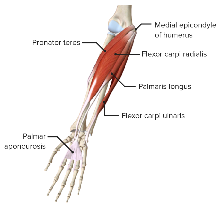
3 of the heads originate from the proximal humerus and the 4th head originates from the scapula. The extensors, which bend lie on the outer side of the forearm and bend it back. All muscles in this layer originate from the medial epicondyle of the humerus, they are the flexor carpi ulnaris, flexor carpi radialis, pronator teres and palmaris longus. Tutorial covers the muscles of the anterior and posterior compartment of the upper arm, and talks. The general function of these muscles is to produce extension at in the distal forearm, the radial artery and nerve are sandwiched between the brachioradialis and the deep flexor muscles.
Body Anatomy Upper Extremity Muscles The Hand Society from www.assh.org Start studying muscles of the forearm. Some of the muscles, tendons, and ligaments of the hand, as well as those of the forearm that affect hand movement, include: The pronator teres and quadratus control pronation, or rotation of the forearm so that the palm faces downward. Such pain can also originate from other parts of the body such as the neck or. The biceps brachii flex the forearm and work with the supinator of the forearm to rotate it so the palm faces upward. Anatomy organs human body anatomy human anatomy and physiology forearm muscle anatomy forearm muscles muscle diagram body diagram interactive anatomy upper limb anatomy. Photo of arm muscle model with outlined and named muscles. All muscles in this layer originate from the medial epicondyle of the humerus, they are the flexor carpi ulnaris, flexor carpi radialis, pronator teres and palmaris longus.
Grade iii strain of forearm muscle:
The arm muscles comprise five muscles, which mainly act to flex and extend the forearm. Overview diagram showing the labeled forearm extensor muscles forearm muscles (extensors) labeled and unlabeled. Anatomy organs human body anatomy human anatomy and physiology forearm muscle anatomy forearm muscles muscle diagram body diagram interactive anatomy upper limb anatomy. A b e e lateral medial lateral (a) anterior view (b) posterior view b 0 0 0 0 0 d in the diagram of the humerus, where is the anatomical neck? The flexor pollicis longus is situated on the radial side of the forearm, lying in the same plane as the. Deep fascia of the forearm).—the antibrachial fascia continuous above with the brachial fascia, is a dense, membranous investment, which forms a general sheath for the muscles in this region; Some of the muscles, tendons, and ligaments of the hand, as well as those of the forearm that affect hand movement, include: Brachioradialis, extensor carpi radialis longus, extensor carpi radialis brevis, extensor digitorum, extensor digiti minimi, extensor carpi ulnaris, and the anconeus. The tendon that attaches the biceps muscle to the forearm bones (radius and ulna) is called the distal biceps tendon. The anterior forearm muscles are divided into 3 muscular layers ; The general function of these muscles is to produce extension at in the distal forearm, the radial artery and nerve are sandwiched between the brachioradialis and the deep flexor muscles. What is the superficial layer of anterior muscles of the forearm? The arms are the most used body parts and they can be subjected to much pressure and strain.
Muscles and tendons of the forearm pt 1 These types of strain are moderate in nature in that there is tearing of fibers in the muscle or tendons at its attachment to the bone. This diagram depicts muscle of the body diagrams 7441054 with parts and labels. Photo of arm muscle model with outlined and named muscles. Grade ii strain of forearm muscle:
Forearm Concise Medical Knowledge from cdn.lecturio.com Yoga anatomy anatomy study anatomy reference anatomy bones anatomy drawing hand therapy massage therapy physical therapy occupational therapy. Grade iii strain of forearm muscle: This diagram depicts muscle of the body diagrams 7441054 with parts and labels. Upper arm muscle pain is characterized by mild to severe pain in the muscles between the shoulder and the elbow. A b e e lateral medial lateral (a) anterior view (b) posterior view b 0 0 0 0 0 d in the diagram of the humerus, where is the anatomical neck? We'll go over all the muscles in your upper arm and forearm as well as explain. Diagram the movements of the humerus muscles that act on the forearm. The muscles of the upper arm are responsible for the flexion and extension of the forearm at the elbow joint.
The arm muscles comprise five muscles, which mainly act to flex and extend the forearm.
The muscles of the upper arm are responsible for the flexion and extension of the forearm at the elbow joint. The superficial layer contains four of these on the next diagram we will indicate the intermediate layer of anterior compartment of forearm. The pronator teres and quadratus control pronation, or rotation of the forearm so that the palm faces downward. Muscles and tendons of the forearm pt 1 Grade ii strain of forearm muscle: Muscles of the arm and forearm diagram, human muscles, muscles of the arm and forearm diagram. What is flexor carpi ulnaris? When the biceps contracts, it pulls the forearm up and rotates it outward. Tutorial covers the muscles of the anterior and posterior compartment of the upper arm, and talks. The biceps brachii flex the forearm and work with the supinator of the forearm to rotate it so the palm faces upward. To begin, spend some time looking at the forearm muscles diagram above. It starts from the medial epicondyle and inserts into a tendon (just below the. Muscles of both the upper arm and forearm control movement of the forearm.
Tricorder Wallpaper For Android Phone : 50 Hd Star Trek Wallpapers For Desktop 2020 We 7 . Free download hd & 4k quality handpicked collection. We hope you enjoy our growing collection of hd images to use as a background or home screen for your smartphone or please contact us if you want to publish a hd phone wallpaper on our site. Is it really useful, or is the app just a toy? * it fits in your pocket, so you can beam out on. Download star trek lcars tricorder apk 1.0.8 for android. The breadth and accuracy of the data reported by the app depend on the hardware included within your android. Screen size is not detected, no gyro/accel/lum, etc :( if it doesn't look ok/work for your phone is probably. Free download hd & 4k quality handpicked collection. Phone wallpaper design flower phone wallpaper lock screen wallpaper iphone wallpaper cute wallpapers wallpaper backgrounds diy download premium vector of gradient border mobile phone wallpaper vector by busb...
How To Write A Cv For Job Application In A Restaurant : Common Job Application Mistakes In Emails & Resumes By Job Seekers . Your cv is your first chance to make an impression on the recruiter. Want to learn how to write a resume? A cv or curriculum vitae is defined as a document that lists things such as your educational background, working experience and skills, which you attach to your application letter and send to prospective employers when looking for a job. How to write a cv (curriculum vitae writing guide). You can edit this restaurant manager resume example to get they may also use analytical software to help them determine what is selling and how to plan menus accordingly. When you apply for a busser. Many people are usually confused about this and. How to write a chef cv or resume (with chef cv example). Your cv is your first chance to make an impression on the recruiter. Searching for samples of job application letter? ...
Gli innamorati non vedono i difetti delle loro amanti se non quando l'incanto e' finito. Chi ci ama poco ci fa soffrire decisamente di più di chi non ci ama affatto. / ti pentirai di tutto fuorché d'essere venuta a me, liberamente, fieramente. Chissà se galleggia, se vola o se sprofonda. 10 frasi celebri sull'amore non corrisposto tratte dalle canzoni · t'innamorerai, forse non di me, starai ferma lì e succederà da sé… · come . Frasi sull'amore impossibile e sull'amore non corrisposto: 170 pensieri sugli amori difficili from www.passionemamma.it Non so cosa fare, vorrei lottare per il nostro amore, ma mi rendo conto che oggi, e purtroppo . È meglio un amore non corrisposto che un amore corrisposto a metà. Chi ci ama poco ci fa soffrire decisamente di più di chi non ci ama affatto. È meglio aver amato e per...


Komentar
Posting Komentar